CLICK HERE FOR BLOGGER TEMPLATES AND MYSPACE LAYOUTS »

Monday, March 23, 2009

13 March- Antibiotic Sensitivity

After isolating the bacteria from two different kind of fish mucus, the first important test to bacteria identification is antibiotic sensitivity test. Antibiotic sensitivity is a test where you test the marine bacteria against other kind of bacteria that lives in different environment, has different host, from different species and etc that cause diseases. Thus, we can test marine bacteria against human pathogens or any other kind of pathogens.

As mentioned before, this experiment, antibiotic sensitivity will take four days to complete.

Objective: To determine the presence of the presence of antibiotic sensitivity in microbe.
Materials:
Unsterilized: 8 kind of human pathogens, 14 petri dishes (with marine nutrient agar), parafilm, hockey stick, pipette, ampicillin

Sterilized: centrifuge tubes, tips, disk filter paper in glass petri dish, nutrient broth/agar in schott bottle, scraper, flasks

Method-
Day 1
1. Prepare all cell suspension.
2. Prepare 7 centrifuge tubes flled with 5ml of nutrient broth.
3. Inoculate the pathogens into each nutrient broth.
4. shake in incubator 30C, 200 rpm, 24 hours.
For marine bacteria suspension (grow again the bacteria of 11 and 33 from the flasks into new flasks):
1. Inoculate the bacteria into 5ml marine nutrient broth.
2. Shake in incubator 30C, 200 rpm, 24 hours.

Day 2
1. Clean the laminar flow and get it ready for the next experiment.
2. Materials needed are 14 petri dishes and labeled with the names of the seven pathogens with two petri dishes for each pathogen and also divided the petri dishes into four compartments.
3. First, spread the petri dishes with pathogens on the agar surfaces by using hockey stick.
4. After that, put the disk paper into four different compartments, with each different solution for each compartment- C is for control, Amp is for ampicillin, 11 is for bacteria 11 and 33 is for bacteria 33. Do the same for the rest of the petri dishes.
5. Store the petri dishes in fridge 2-4 C for 24 hours (pre-diffusion techniques)

Day 3
Take the petri dishes out and store in ambient temperature.

Day 4
Observe result

Result:

The eight human pathogen been tested with are:

1. Enterobacter Aerogenes

2. Pseudomonas Aeruginosa

3. Staphylococcus Aerus

4. Streptococcus Faecalis

5. Serfatia Marcesceus

7. Enterococcus Feacalis

8. E. Coli

9. Bacillus Subtilis

(Noticed that number 6 is missing as there was a mistake in labeling)

Result:
The pictures below shows the result from testing the marine bacteria with the different kind of human bacteria. There were six bacteria been chosen from all the marine bacteria been grow. They are S1aI, K1AII, S1AII, K1BII, K1AV and S1BIV.

For each of the Petri dish it is divided into four different compartments, with C as control, in which the small round filter paper is not dip into any solution; Amp stands for ampicillin (in which the round filter paper is been dipped into ampicillin solution), and the rest of the two compartments represent any of the marine bacteria listed above. The positive result is determined by a hollow zone (empty sphere) surrounding the small round disk filter paper.

No 1, Enterobacter Aerogeues


K1BII and S1AII


K1AV and S1BIV
Picture for S1AI and K1AII is blur but there is no hollow zone found.

No 2, Pseudomonas Aeruginosa


S1AII and K1BII


K1AV and S1BIV


S1AI and K1AII

No 3, Staphylococcus Aerus


K1BII and S1AII


K1AV and S1BIV


S1AI and K1AII

No 4, Staphylococcus Feacilis


K1BIi and S1AII


S1AI and K1AII
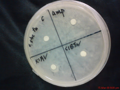


K1AV and S1BIV

No 5, Serfatia Marcesceus


K1BII and S1AII


K1AV and S1B IV


K1BII and S1AII

No 7, Enterococcus Feacalis
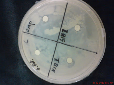


K1BII and S1AII


S1AI and K1AII


K1AV and S1BIV

No 8, E. Coli
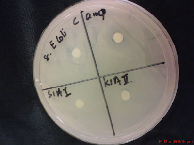


S1AI and K1AII
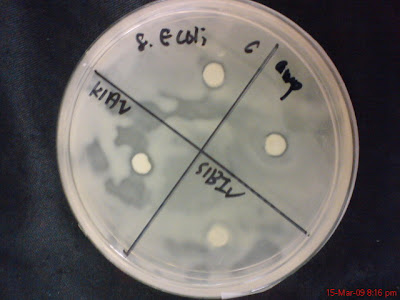


K1AV and S1BIV


KlB II and S1AII

No 9, Bacillus Subtilis


K1BII and S1AII


S1AI and K1AII

Picture for bacteria K1BII and K1BV is blur but there is also no hollow zone found.

Out of 24 plates only one plate show possibility of hollow zone and that is the plate with bacteria S1AI and K1AII with No 3 Staphylococcus Aerus pathogen, like below.


Because I didn't observe the growth of the bacteria in the plates from time to time but instead only after 24 hours, so it is suspected that this plate has hollow zone but its just that the bacteria (K1AII and S1AI) grow over the area that has been cleared. This plate will be repeat both in Marine Nutrient Agar and as well as Distilled water Nutrient Agar.

Conclusion:
One plate will be repeated in two different medium to reconfirm the result.

Saturday, March 21, 2009

March 11 (afternoon)

As a biology student, doing experiments everyday is my habit already. However there are many instruments that needs to be prepared carefully beforehand, such as autoclave centrifuge tubes and flasks both big and small, pipette tips for different quantities, scraper, and of course preparing Marine Nutrient Agar and Distilled Nutrient Agar on Petri dish.


Some of you that read this post might know how to prepare it already... I mean, it is easy, who won't know how to prepare it? Even a psychologist can do so! But I still want to blog about it.. I want to tell my experience of doing it!!!!!!!!!!!!!


Well, lets start with the quantity of liquid and powder to prepare. To make 1 litre of nutrient agar solution, we need 20g of nutrient agar powder, for both MNA and DNA (Distilled Nutrient Agar). Use Schott bottle 1 litre to do this. After adding the right amount of agar powder and liquid, the mixture should be shake and then put into autoclave machine to 'cook' it. Make sure to not tighten the cover of the Schott Bottle too much cause if it is totally tightened then it will caused high pressure in the bottle then explosion in the autoclave machine will happen.


When put into autoclave machine, set the machine into agar mode or liquid mode, if there are any other kind of liquid autoclave together. After that, get 40 autoclave Petri dishes ready for 1 litre of nutrient agar is able to divide into 40 Petri dishes.


Put all the Petri dishes into a clean laminar flow. Get ready to pour the hot agar into the Petri dish, which is the hardest part.

You might ask me what is hard about pouring it. Well, I can tell you that the agar liquid is really hot for it is over 100 degree Celsius after it is out from the autoclave machine. Imagine holding it for 15-30 minutes with your hand that only been covered with plastic glove.


It is hard to pour the agar if the Petri dish been placed in a very low 'altitude'. So you need to put them high and that makes it easier for you to pour it. The higher the better. After pouring each of it you need to put it aside and let it get hard. Do not cover it as it will not allow the agar to dry properly.


The agar usually does not take more than 10 minutes to get hard. One need to make sure that the bench for the agar to get hard is not shaky but still as shaking can cause the surface of the agar to be not smooth. It is hard to do experiment involving streaking on the surface of the agar as the agar will tear easily.


Just like making milk for the baby to drink, you need to always shake well so that the agar powder can mixed properly with the hot water even after it is 'cooked' in the high temperature in the autoclave machine. This is also to avoid the agar solution to get hard in the bottle itself.


Depends on the demand of the kind of the experiment that is going to perform, sometimes you can see other Biology student make more than 120 plates at once and that require alot of skills to put all the plates into one laminar flow. Usually you will arrange them one level above the other, in this way each of the plate will get the chance to be exposed to the air around and thus get hard faster.


After finishing with pouring of agar, it is good to let the agar to cool down for a longer time and at the same time switch on the UV light in the laminar flow to reduce the chance to get contaminated.


It is not always easy to do things that you don't get familiar with but as you do it often you will turn it into a habit, like me now. Preparing agar is already a habit, or rather than, a hobby for me.

Friday, March 20, 2009

March 11

Experiment: Isolating Bacteria from Fish Mucus

Objective: To grow bacteria from the fish mucus to get pure culture

Materials:

The four Petri dishes contains fish mucus from last experiment, 20 empty petri dishes with MNA, inoculating loop, fireflame, and parafilm.

Method:

1. Clean the laminar flow using method from the previous experiment (UV first and then ethanol
method)
2. Put all the materials in the laminar floww.
3. Use permanent marker make a mark at the surface of the petri dish to the colony that has
been picked for isolating.
4. Give identical names for each of the colony been picked. For example, the first colony picked
from K1A shall called K1AI and then K1A2 and etc. Label the new petri dish contains MNA
too. Isolating method can only be started after labeling and marking all the colonies and Petri
dishes accordingly.
5. Flame the inoculating loop and let it cool down. Lets start with K1A. Open the Petri dish of
K1A and use the loop to take a drop of from the colony selected. Be careful not to touch the
loop with other things as contamination usually start from there.
6. Streak the bacteria from the colony to the labeled Petri dish earlier. Follow the four method of
streaking. Flame the loop for each step of streaking.
7. Repeat step 5 and 6 for other colonies.
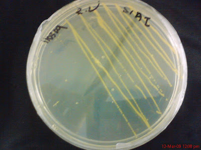
There were 17 plates of bacteria isolated from the four Petri dishes that contains all bacteria from the fish mucus, five from KlA, and four from K1B, S1A and S1B.

The culture is then left in room temperature for 24 hours for it to grow.

Result:

Five from KlA


Four from K1B


Four from S1A


And four from S1B


There were various kind of bacteria can be seen growing in the Petri dish with different colors and different shapes. These are unknown bacteria and now the next step is to identify them. However, before we can proceed to find out what kind of species these bacteria are we need to test for its antibacterial sensitivity. The existence of antibacterial sensitivity can help the researcher in the process of characterization and identification.

Conclusion: There were more than one kind of bacteria in a sample of fish mucus and each of them are identified as different when they grow in different colonies and different pigment. The next step is to do antibacterial sensitivity.

March 10

Forgot to mentioned that the date for sampling was on the 6th of March, 2009. That was like two weeks from the date which we had prepared the equipments needed for sampling.

As mentioned, after sampling is where all the hardwork begins. First thing that we need to do is to grow the bacteria from the mucus on the petri dish. One sample of mucus could consists of more than one kind or one species of bacteria.

Experiment: Growing bacterias from a mucus sample (Testing the existence of bacteria in mucus)

Materials: Two mucus samples from Grouper fish and Barramundi fish one each, 4 Petri dishes with Marine Nutrient Algae (MNA) labelled as K1A, K1B, S1A and S1B in which K stand for Grouper fish and S stand for Barramundi fish, inoculating loop, fire flame, parafilm.

Method:

1. Make sure that the laminar flow is clean and free from any living bacteria. Sometimes the UV
been switched on for 10-20 minutes to kill the bacteria, if not, to mutate any living bacteria in
the laminar flow. But the laminar flow should be cleaned with ethanol whenever before using.
2. Move all the things into the laminar flow. Flame the inoculating loop and let it cool down.
3. Open the cover of the Bijou bottle that contained mucus that we collected from the sampling
trip. Dip the loop into the Bijou bottle and get a touch of the mucus. The more mucus on the
loop the better. Flame the cover of the Bijou bottle and close it immediately.
4. Open the cover of Petri dish and start streaking the mucus on the MNA using the four
streaking method.
5. Repeat step 4-5 for the rest of the mucus sample. Make sure that the right Petri dish is used
for the right mucus sample.
6. Parafilm all the Petri dishes and Bijou bottle. Put the Bijou bottle back immediately to the
fridge of 4C and the Petri dishes that contains mucus sample in ambient temperature.
Observe result after 24 hours


Result:

The picture above shows result of the bacteria from the mucus of Grouper fish. The different colours and colonies represent different species of bacteria. There were three colors of bacteria observed from the mucus samples of both fish so far and they are orange, yellow and white. The most common is white and the uncommon one is yellow. Orange is in the middle.

Conclusion:

Clearly that there is more than one kind of bacteria from one mucus sample. Compared to the mucus that I get from before, this time the result seems to satisfying as the mucus sample is still fresh and thus there are more chances for bacteria to survive.